Brain Scan Might Someday Spot Autism
 A kind of brain imaging that measures the circuitry of brain connections may someday be used to diagnose autism, new research suggests.
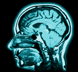
Researchers at McLean Hospital in Boston and the University of Utah used MRIs to analyze the microscopic fiber structures that create the brain circuitry in 30 males aged 8 to 26 with high-functioning autism and 30 males without autism.
Males with autism showed differences in the white matter circuitry in two regions of the brain's temporal lobe: the superior temporal gyrus and the temporal stem. Those parts are involved with language, emotion and social skills, according to the researchers.